All Children’s Hospital Los Angeles locations are open.
Wildfire Support Line for Current Patients, Families and Team Members:
323-361-1121 (no texts)
8 a.m. - 7 p.m.

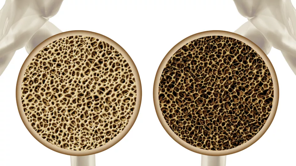
Patients with acute lymphoblastic leukemia are at increased risk for bone fractures due to bone loss resulting from treatment. This decrease in bone density (as illustrated here) can occur as soon as the first month of treatment according to new research out of Children’s Hospital Los Angeles. Image courtesy of Shutterstock.
Investigators at Children’s Hospital Los Angeles have found that significant bone loss – a side effect of chemotherapy for acute lymphoblastic leukemia (ALL) – occurs during the first month of treatment, far earlier than previously assumed. The study was published online in the journal Bone on February 4.
ALL is the most common pediatric cancer. Forty years ago, only one in five children survived this disease. Today with the development of powerful chemotherapies, over 90 percent of patients can expect to be cured. Unfortunately, there are some significant side effects to these life-saving therapies, including loss of bone density resulting in an increased risk for bone fractures during and even after therapy. Previous studies to determine the changes to bone density during ALL therapy had focused on the cumulative effects of chemotherapy after months or even years of treatment.
“In clinic, we would see patients with fractures and vertebral compression during the very first few weeks of treatment,” said Etan Orgel, MD, MS, first author on the study and an attending physician in the Survivorship & Supportive Care Program at the Children’s Center for Cancer and Blood Diseases at CHLA. “But we were unaware of any study that specifically examined bone before chemotherapy and immediately after the first 30 days of treatment – which would allow us to understand the impact of this early treatment phase.”
In a prospective study in newly diagnosed patients 10 to 21 years of age, the investigators explored leukemia-related changes to bone at diagnosis, and then the subsequent effects of the first – or induction – phase of chemotherapy. Using quantitative computerized tomography (QCT) – a newer technique more accurate for use in growing bone – they determined that leukemia did not dramatically alter the properties of bone before chemotherapy (in comparison to similar age- and sex-matched control patients).
During the 30-day induction phase, however, bone mineral density of the lower spine decreased by more than 25 percent with significant thinning of the dense cortex occurring in the bones of the leg. To help clinicians relate to these findings, the team also measured bone mineral density using the older but more widely available technique of dual-energy x-ray absorptiometry (DXA) and found that it underestimated these changes as compared to QCT measurements.
“Now that we know how soon bone toxicity occurs, we need to re-evaluate our approaches to managing these changes and focus research efforts on new ways to mitigate this common – yet significant – adverse effect,” said Steven Mittelman, MD, PhD, principal investigator at The Saban Research Institute of Children’s Hospital Los Angeles and senior author on the study.
Funding was provided in part by the Leukemia and Lymphoma Society, the Southern California Clinical and Translational Science Institute and The Eunice Kennedy Shriver National Institute of Child Health and Human Development.