Preterm Birth Can "Hardwire” Brain Abnormalities Persisting into Adulthood
Preterm birth is linked to lasting physical changes in a child’s brain. Each year, around 15 million infants are born prematurely, placing them at high risk for adverse neurodevelopmental outcomes. But whether the brain tissue abnormalities that accompany preterm birth persist into adulthood and are linked with long-term cognitive or psychiatric outcomes have been open questions.

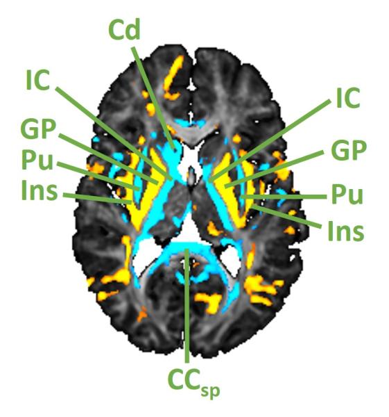
Recently, a team led by Bradley Peterson, MD, published the results of a two-decade study in the Journal of Child Psychology and Psychiatry that began to address these issues. The team followed 180 Norwegian preterm and full-term infants over roughly two decades and assessed participants using cognitive measures and psychiatric diagnoses. They scanned participants' brains using diffusion tensor imaging (DTI) at 18 to 19 years of age.
What the scans revealed
The DTI scans found widespread microtissue disturbances in the organization of the preterm group’s brains. Tissue abnormalities were worse if a child had a younger gestational age at birth as well as complications involving infection or inflammation.
“The findings were very powerful,” says Dr. Peterson, Chief of the Division of Psychiatry and Co-Director of the Behavioral Health Institute at Children’s Hospital Los Angeles and lead author of the paper. “They showed prominent abnormalities in deep white matter tracts throughout the brain.” The abnormalities likely represent disordered myelination in the axons or nerve cells that organize the brain’s communications between different regions. Abnormalities were observed in basal ganglia nuclei and thalamus deep in the brain which are important in executive functioning; planning and regulating, thought processes, emotions, and behavior.

“Our findings show very clearly that these abnormalities don’t resolve by adulthood,” says Dr. Peterson. Imaging also uncovered abnormalities in the brain’s gray matter. “Many kids born prematurely have problems with this," says Dr. Peterson. “It seems that these brain disturbances in most people born prematurely are hardwired from the beginning of life, beginning even in utero. They come into the world with these brain differences, and those differences persist into adult life.”
Clues to causes
Prior research in animal models has shown that inflammation and oxidative stress likely contribute to these brain differences, particularly in the second trimester of pregnancy when developing cells in the white matter of the brain are most vulnerable. These stresses, either in the mother or the fetus—are toxic to the precursor cells that form myelin in the brain’s white matter—and can cause dysfunctions that persist into adulthood.
Multiple studies by different groups are underway—both in animal models and in human clinical trials— to investigate dietary supplementation and novel compounds that could potentially reduce the effects of inflammation and oxidative stress. “In my opinion, the million-dollar question is how to mitigate those brain effects of premature birth,” says Dr. Peterson.


