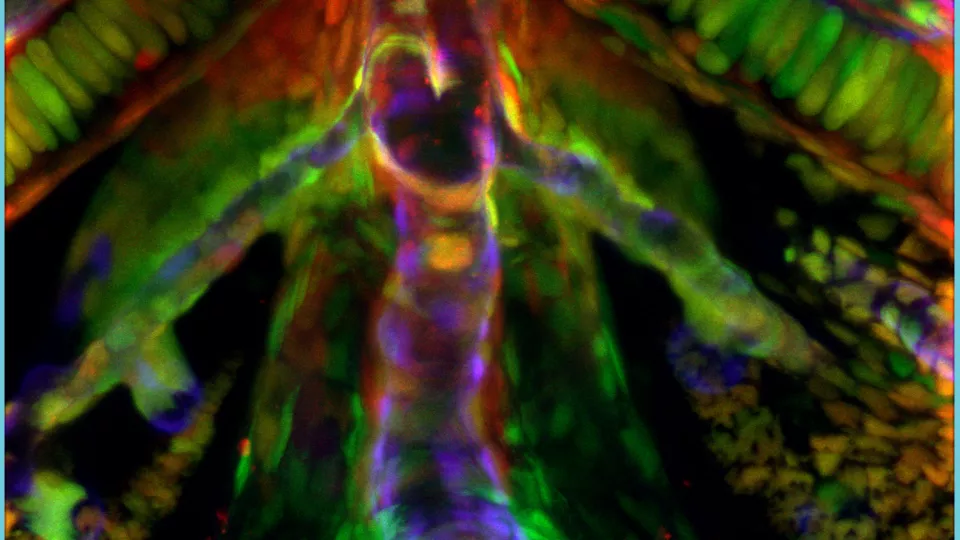
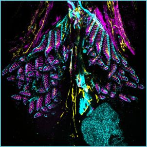
An image produced in the Cellular Imaging Core, showing the ventral aorta (an artery through which blood is pumped from the heart to the gills) of a live zebrafish (photo by Yuhan Sun, PhD)
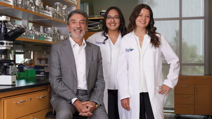
How Do You Take a Picture of a Cell? CHLA’s Cellular Imaging Core Has the Answers
They’re colorful. They’re fluorescent. And they wouldn’t look out of place hanging in a modern art museum.
They are the photos captured by the Cellular Imaging Core, one of 10 research cores at The Saban Research Institute of Children’s Hospital Los Angeles. Each core functions as a centralized resource for CHLA’s researchers, offering access to shared equipment and advanced technology services. Access to these cores eliminates the need for dozens of specialized tools to be obtained for every single lab at CHLA. Instead, investigators, trainees and staff from across CHLA can access equipment and services and receive advanced training for equipment use.
The Cellular Imaging Core provides state-of-the-art light microscopes and digital imaging equipment to researchers, enabling them to obtain and analyze images of single cells and tissue samples. This advanced imaging is a key part of The Saban Research Institute’s many biomedical research projects.
By isolating fluorescent proteins and utilizing genetics and new methods for localizing proteins and expressed genes, the Core uses a variety of laser-equipped microscopes to image the complex molecular architecture of cells from healthy and diseased tissues. Services provided also include training in the use of digital imaging technology and the analysis of images to generate data.
Twelve images produced in the Core by CHLA researchers were entered into an Image Contest hosted by G. Esteban Fernandez, PhD, Director of the Cellular Imaging Core, as part of The Saban Research Institute Science Day 2024. This annual contest is open to all CHLA researchers. Science Day attendees and a panel of judges vote for their favorite images, and prizes are awarded to the winners. For Dr. Fernandez, who created the Image Contest, seeing the enthusiasm for scientific images at CHLA represents the culmination of his own decades-long passion.
A dream job
“There’s something about the physics and the biology of imaging,” Dr. Fernandez says. “It’s highly satisfying.” He became interested in imaging while earning his undergraduate degree in engineering, recognizing that it combined his love of science with his interest in developing technologies.
In graduate school, he applied advanced microscopy to his thesis project and learned that there were many ways to process and analyze images using digital technology. “A lot could be done with the images after they were produced, and I knew how to software code from my undergraduate days. So it was a lot of fun to digitally work with images.”
From then on, Dr. Fernandez was hooked. “I decided to focus on imaging as a career,” he says.

He soon learned about scientific imaging core facilities at biomedical research institutions, which feature specialized imaging equipment and provide expert services. “I thought, ‘That sounds like a dream job,’” Dr. Fernandez says.
He first worked at a core facility in Missouri before shifting to his present role in the Cellular Imaging Core at CHLA, where he has been for the past 15 years. During this time, he has built the Core up from just a few standard microscopes and one advanced scope to an array of the latest-generation microscopes, with added functionality that he has spent years fine-tuning.
A large part of Dr. Fernandez’s role in the Cellular Imaging Core is keeping up with technological advances. “As new technology comes into the marketplace, I need to keep abreast of that and also have a have a sense for the projects that researchers are doing at CHLA and kind of merge the two together,” he explains.
He uses this expertise to train CHLA scientists to independently produce images for their own research purposes using the Core’s instruments.
The Cellular Imaging Core has grown in both equipment and staff. When Dr. Fernandez added a sophisticated microscope capable of 2 photon and live cell imaging, he hired Senior Research Associate David Koos, PhD, who was particularly experienced in those imaging techniques. “He’s like me,” Dr. Fernandez says. “This is his dream job.”
This shared passion extends to the Image Contest, now an annual tradition. He and Dr. Koos are both on the judging panel for the contest, along with Paolo Neviani, PhD, Stephan Erberich, PhD and Long Hung, all leaders of research cores and The Saban Research Institute Data Science team.
“I thought it could be a way to increase visibility for the Core,” Dr. Fernandez explains. “Maybe people know that we exist, but they don’t know exactly what kinds of images can be taken or what techniques we can use. And l hoped it would draw people to science because they can have fun just looking at the images and learning.”
Award-winning images
Toward the end of Science Day, Dr. Fernandez announced the 2024 Image Contest award recipients (who were given professionally printed and mounted versions of their images, as well as a small cash prize):
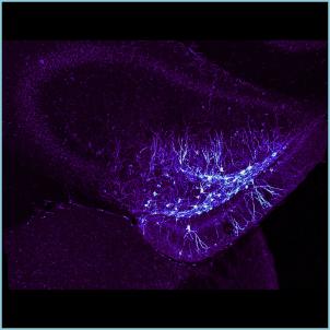
- Judges’ Runner-up: Doctoral student Laura Korobkova, in the lab of Brian Dias, PhD, for an image of neurons in the dentate gyrus of a mouse hippocampus (a region in a mouse’s brain), produced using an optimal imaging technique called confocal microscopy on the Cellular Imaging Core’s Leica STELLARIS 5 microscope.
- Judges’ Favorite: Postdoctoral fellow Yuhan Sun, PhD, in the lab of Ching-Ling (Ellen) Lien, PhD, for an image of the ventral aorta (an artery through which blood is pumped from the heart to the gills) of a live zebrafish, produced using confocal microscopy on the Cellular Imaging Core’s Zeiss LSM 710 microscope.
- Audience Runner-up: Doctoral student Hui Yu (Elliot) Chen, in Dr. Lien’s lab for an image of a zebrafish gill and heart, produced using confocal microscopy on the Cellular Imaging Core’s Leica STELLARIS 5 microscope.
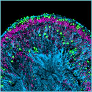
- Audience Favorite: Doctoral student Jinlun (Jay) Bai, in the lab of David Cobrinik, MD, PhD for an image of a human retinal organoid (a 3-D tissue made from retinal stem cells in the eye), produced using confocal microscopy on the Cellular Imaging Core’s Leica STELLARIS 5 microscope.
Dr. Fernandez sees the contest as both educational and entertaining. “Humans are very visual creatures,” he says. “So if you have tons of data showing that a phenomenon is occurring, I think seeing it in a picture is most compelling.”