Small Animal Imaging Core Equipment and Software
Equipment
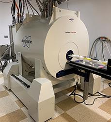
11.7 T BioSpec uMRI (Bruker) 2020
A multipurpose high field MR scanner for both magnetic resonance imaging(MRI) and magnetic resonance spectroscopy(MRS) for preclinical, pharmaceutical, and fundamental research. The instrument is based on the AVANCE NEO MRI scanner architecture, consisting of 11.7T, 11cm Bore US/R magnet, Faraday RF-Shielding Cabinet, Main Gradient and Shim, MRI RF Coil, MRI Cryo Coil, 1H Transmit and Receive Channel.
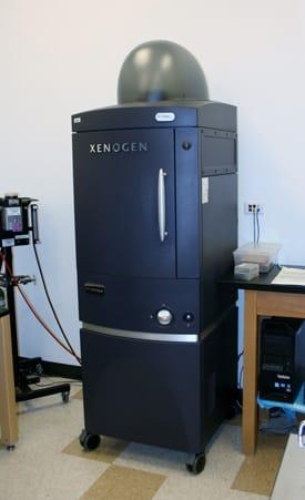
IVIS 200 Bioluminescence/Fluorescence (Perkin-Elmer, former Xenogen) 2013
In vivo imaging technology platform, that allows non-invasive visualization and tracking of cellular and genetic activity within a living organism in real time. This equipment is work-horse with high throughput capacity. An optimized set of high efficiency filters and spectral un-mixing algorithms lets you take full advantage of bioluminescent and fluorescent reporters across the blue to near infrared wavelength region. It also offers single-view 3D tomography for both fluorescent and bioluminescent reporters that can be analyzed in an anatomical context using Digital Mouse Atlas.
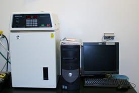
Faxitron MX-20
The industry standard in cabinet X-ray systems for point-of-care specimen/small rodent radiography, specified in bone studies and implants.
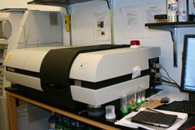
SkyScan1172 HighRes uCT (Bruker)
High-resolution desktop X-ray uCT system for small samples. The flexible geometry of the scanner is particularly advantageous over intermediate resolution levels. Good for biological tissue structural studies.
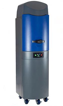
Lago X (2022)
Free standing Optical Imaging System for Small Animal Imaging is an exceptional tool for whole-body applications, with its exceptional reproducibility, signal stability and camera sensitivity. LAGO X Bioluminescence Imaging instrument offers high throughput imaging for BLI, FLI, and X-Ray. The LAGO X provides Absolute Calibration, -90C Air Cooled CCD Camera, LED Fluorescence excitation. It also has an X-ray imaging mode that can be overlaid for anatomical reference of the acquired signals. The Fluorescence illumination is LED based, with 14 excitation wavelengths and 20 emission filters. The mouse capacity of the imaging chamber is 10 mice on a 25cm x 25cm field of view. The AURA software used for the analysis of the .ami image files obtained in the Lago X is free to all users. PC and Mac options available. Download at spectralinvivo.com/software.
Imaging Data Analysis
The Research Imaging Core is equipped with a wide variety of commercial tool sets for imaging data analysis and visualization.
CTAn, CTVol - Bruker's data analysis and visualization tool suite
Analysis - Olympus's image analysis software
Amira - FEI's platform for 3D and 4D data visualization, processing and analysis
Fiji (ImageJ) - A NIH image processing tool
Lightwave 3D - NewTek's 3D computer graphics software
Imaris - 3D and 4D Real-Time Interactive Data Visualization Platform
Living image - In Vivo Imaging Software
Arivis 4D - Data visualization Platform
Analyze Direct – Image processing software
MATLAB – computing environment and proprietary programming
Visual Studio - Interactive developmental environment for programming