CHLA Researcher Uses Low-Field MRI to Assess Lung Capacity in Children With Single Ventricle Hearts
As surgical techniques improve, more children born with single-ventricle heart disease are surviving to middle age, even without a heart transplant. But with only a single ventricle, the heart cannot pump enough oxygen-rich blood to the lungs. “We are examining this mismatch,” says Eamon Doyle, PhD, Data Engineer, Data Science and AI at Children’s Hospital Los Angeles and Assistant Professor of Clinical Radiology, Keck School of Medicine of USC.
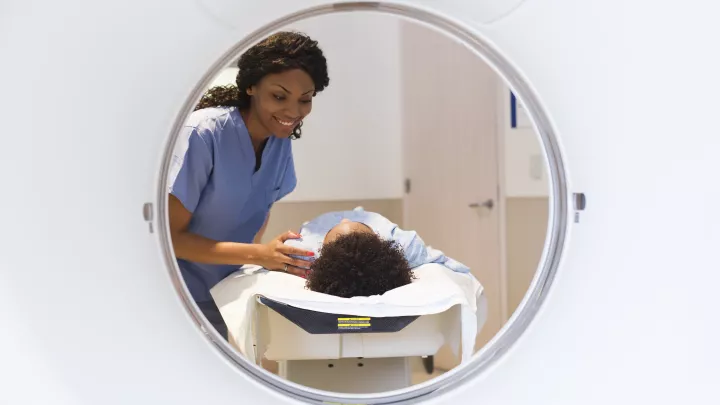
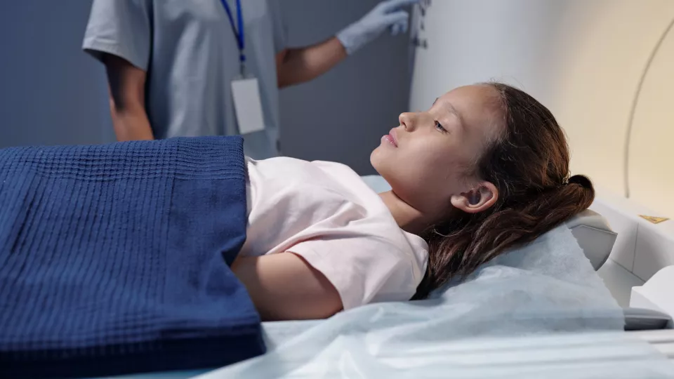
Under a $50,000 1-year grant from the Additional Ventures Foundation and a Cardiac Imaging Suite Pilot Award, Dr. Doyle will use novel magnetic resonance imaging (MRI) technologies to scan the hearts and lungs of children between 12-18 years old born with single ventricle hearts, as well as those of healthy control patients. His goal is to assess methods of determining blood flow, ventilation patterns, and oxygen perfusion in the lungs.

“We hope this data will lead to therapeutic targets or allow us to assess the treatment efficacy and improve our understanding of blood flow,” says Dr. Doyle. “There may even be opportunities for 3D modeling of surgical approaches to figure out why some patients do better than others, or if there's a preferred surgical approach that will have better long-term outcomes.”
Project co-investigators include Heart Institute Attending Physician and researcher Jon Detterich, MD, MR Physicist Xin Maio, PhD, and John Wood, MD, PhD, Director, Cardiovascular MRI at the Heart Institute at CHLA, and pulmonologist Roberta Kato, MD.
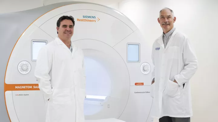
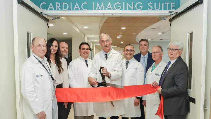
The Cardiac Imaging Suite—a unique imaging toolset
Dr. Doyle is using two state-of-the-art MRI systems in CHLA’s new Cardiac Imaging Suite: a cardiology-specific Siemens Sola 1.5 Tesla MRI machine, and the Siemens FreeMax 0.55 Tesla MRI Machine, a new low-field, high-resolution magnet that reduces certain image artifacts, making imaging the heart and lungs easier because the signals don't disappear as quickly as they do at higher field strengths. “It gives us more time to acquire data and enables more contrast in the lungs than other systems,” Dr. Doyle says. “We are the first children's hospital in the nation with the FreeMax installed, which gives us a huge advantage.”