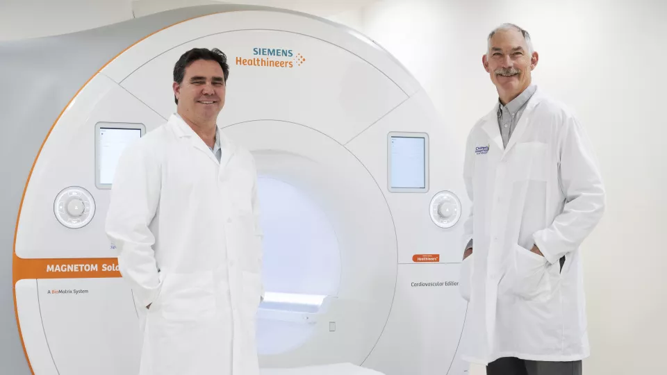
Left to right: Jon Detterich, MD, and John Wood, MD, PhD, in the new Cardiac Imaging Suite at CHLA
Q&A: How CHLA’s New Low-Field MRI Could Transform Pediatric Imaging
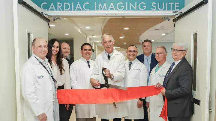
On Dec. 9, Children’s Hospital Los Angeles unveiled its new, FDA-approved low-field MRI machine—the first in the U.S. at a pediatric center. Cardiologist Jon Detterich, MD, completed the first scan on the Siemens FreeMax 0.55 Tesla MRI machine, which is part of CHLA’s newly opened Cardiac Imaging Suite. The new suite also houses a state-of-the-art 1.5T MRI.
What benefits does low-field MRI have for pediatric patients? What tests are possible now, and what does the future hold? International MRI expert John Wood, MD, PhD, Director of Cardiovascular MRI in the Heart Institute at Children’s Hospital Los Angeles, shares what’s happening—and why this new technology is a game-changer for pediatric imaging.
What are the advantages of low-field MRI?
Low-field MRI works particularly well for any body structure that moves or is near air. With a stronger magnet, you have a stronger signal, but movement or air can cause distortion in the image. Because the 0.55T has a weaker signal, you don’t get that distortion.
This means that low-field MRI is ideal for imaging structures like the heart. It allows imaging of the lungs and the GI tract, which we can’t do with high-field magnets. And it’s more suited for imaging a fetus, which is constantly moving around the womb.
The machine is also significantly quieter than a high-field MRI and much more spacious, so it’s more comfortable for patients. And because it accommodates motion, the patient doesn’t have to be perfectly still. For kids, that means we may not have to use general anesthesia for the scan, which is huge.
How is CHLA using this machine right now?
We are already using it to get excellent, diagnostic-quality images of the heart, in this quieter, much more spacious environment.
Low-field MRI is also excellent for patients who have metal implants. We are scanning children and teens who have pacemakers, for example. Our neurosurgeons and orthopedic surgeons are also interested in referring their patients with metal implants to this machine.
Can low-field MRI be used for real-time imaging?
We are not yet using this clinically, but we have been partnering with Krishna Nayak, PhD, and his team in the USC Viterbi School of Engineering, to develop real-time imaging capabilities.
For example, two years ago, our group was the first in the world to perform real-time imaging of a human fetal heart—without the need for cardiac synchronization—using low-field MRI. You could see the fetal heart beating, real-time. That had never been done with MRI.
Our ultimate goal is to use this magnet to guide procedures such as cardiac catheterizations. If we could use low-field MRI instead of X-ray, that’s a big deal—especially for children, where we really want to limit radiation exposure.
What non-cardiac applications is the team working on?
One of the big potential applications for pediatrics is lung imaging. Right now, from an imaging perspective, we don’t have a good way to monitor progression of lung disease in babies and children. You can’t take multiple CT scans in children who have cystic fibrosis or other lung disease. There’s too much risk from radiation.
Currently we have a whole team studying low-field MRI for children with lung disease—including children with Fontan circulations who have lung issues. Another group is focusing on swallowing studies and speech.
It will take time to develop these techniques and translate them to the clinical environment. But that is where we are headed.
How do you see the future of low-field MRI in pediatric imaging?
First, let me be clear: There will always be a role for standard 1.5T cardiac imaging. It’s really good. It’s what we use day in and day out, hundreds of times a year.
That said, I do believe low-field MRI will become an important tool in the pediatric imaging toolkit. I think it’s going to become the mainstay of imaging for all those areas where 1.5T doesn’t work as well, such as the lungs and abdomen and certain areas of the heart.
It’s going to take time to get there. But it’s tremendously exciting to be first in the U.S. with this new technology. It’s going to open up a world of possibilities that can improve care for children.