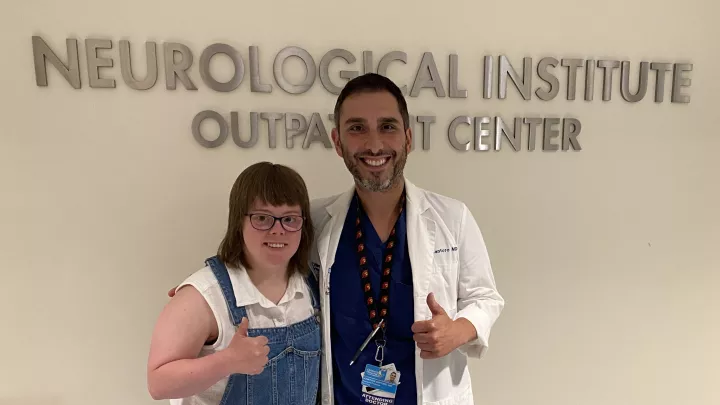
Jonathan D. Santoro, MD
Breakthrough Study Led by CHLA Neurologists Links Blood-Brain Barrier Dysfunction and Inflammation With Down Syndrome Regression Disorder
A new research study led by Jonathan D. Santoro, MD, Director of the Neuroimmunology Program at Children’s Hospital Los Angeles, shows evidence of dysfunction of the blood-brain barrier and inflammation in the central nervous system in individuals with Down syndrome Regression Disorder (DSRD).
The new study, “Evidence of blood–brain barrier dysfunction and CSF immunoglobulin synthesis in Down Syndrome Regression Disorder,” was funded by the National Institutes of Health (NIH) and was published in the Annals of Clinical and Translational Neurology on Feb. 25, 2025. It was led by Dr. Santoro in collaboration with Saba Jafarpour, MD, Natalie K. Boyd, Benjamin N. Vogel, Lina Nguyen, and Lilia Kazerooni, from CHLA’s Neurological Institute. The team collaborated closely with the Linda Crnic Institute at the University of Colorado and its director, Dr. Joaquin Espinosa.
“This is a huge missing piece in the puzzle of what we know about Down syndrome Regression Disorder,” says Dr. Santoro.

Leading DSRD research efforts
Over the past several years, Dr. Santoro, Dr. Jafarpour, and their team in the Strategic Therapies for Overcoming Reactive iMmunology (STORM) Lab have been leading several research efforts and developing new treatments for DSRD. This rare, yet increasingly diagnosed condition causes a rapid decline in young people with Down syndrome. High-functioning individuals will abruptly lose the ability to communicate, feed themselves, sleep, get dressed, or use the bathroom. Some individuals with the disorder become immobile and catatonic.
Though DSRD was first described in a paper in 1946, it was not extensively researched since many assumed it was a psychiatric condition or potentially early-onset Alzheimer’s disease, which are all well described in this population. But Dr. Santoro’s team began seeing patients with DSRD and investigated it further. Their early research identified inflammatory markers in patients’ cerebrospinal fluid indicating that DSRD could in fact be an inflammatory condition affecting the brain.
“This was the aha moment,” says Dr. Santoro.
This discovery informed Dr. Santoro’s treatment approach for DSRD, and he began administering high-dose steroids, as well as an immune therapy known as intravenous immunoglobin (IVIG). This approach proved highly effective, enabling patients to regain the ability to walk, run and communicate.
But clinical investigations have identified abnormalities in cerebrospinal fluid that indicate neuroinflammation in only a fraction of patients with DSRD, which didn’t line up with the high success rate of immunotherapy among this population. Dr. Santoro’s team and their collaborators at the University of Colorado continued their in-depth research.
New evidence of DSRD as an inflammatory condition
This latest study finds clear evidence that DSRD is indeed an inflammatory condition. It also connects DSRD with dysfunction of the blood-brain barrier, a layer of cells that form a membrane between the blood and the brain to filter out harmful substances. “The blood-brain-barrier is critical for keeping the immune system out of the brain,” Dr. Santoro explains. “Any disruption could be enough to cause neurologic disease.”
The new study involved samples of cerebrospinal fluid from three different patient populations: individuals with DSRD, individuals with a known neuroimmunologic or neuroinflammatory condition, such as multiple sclerosis or autoimmune encephalitis, and a neurotypical, non-inflammatory control group.
Proteomic profiling of samples from these individuals allowed the researchers to study the proteins found. Metabolomic profiling identified the metabolites—the molecules created when food, drugs, chemicals, or tissue is broken down—and enabled the researchers to identify the functional status of cells in the body. And immune marker profiling analyzed the specific molecules expressed by immune cells in the samples.
Indications of inflammation and blood-brain barrier dysfunction
The data showed heightened dysregulation in proteomics signatures in DSRD and neuroinflammatory patient samples when compared with the healthy control patient samples, specifically upregulation of several immunoglobulin sequences that indicate neuroinflammation. Additionally, the DSRD patient samples displayed significant upregulation of erythrocyte proteins and liver-derived plasma proteins, which point to poor integrity of the blood-brain barrier. The immune marker profile of the DSRD patient samples was also similar to various other neuroimmunological conditions.
Based upon these results, Dr. Santoro and his team determined that the cerebrospinal fluid of individuals with DSRD has proteomic and metabolic signatures that are consistent with both neuroinflammation and increased permeability of the blood-brain barrier.
“The proteomic abnormalities found in this study confirm what we have seen in clinical practice – the immune system is a major player in DSRD,” Dr. Santoro says.
The cerebrospinal fluid of DSRD patients is also more comparable to that of patients with neuroinflammatory disorders than to healthy control patients, suggesting a potential immune-related cause of DSRD which could inform future treatment strategies.
“The implications of these findings are profound and will hopefully serve as the next step for finding the cause of DSRD” Dr. Santoro explains.
This research has led to a Phase IIb clinical trial assessing the safety and efficacy of immunotherapy in individuals with DSRD. This clinical trial, performed with the Linda Crnic Institute at the University of Colorado, is the first of its kind in the treatment of DSRD.
As Dr. Santoro says, “We’ve come a long way but have more work to do!”